|
|
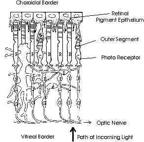
Figure 2. The adult vertebrate retina. In this representation, light enters from the bottom of the diagram (i.e., at the vitreal border). The main cell types are rods (R), cones (C), horizontal cells (H), bipolar cells (B), amacrine cells (A), and ganglion cells (G). At the top of the figure is the RPE. Its finger-like cellular processes interact with the photoreceptors. (Figure redrawn from R. Adler and D. Farber, The Retina [New York: Academic Press, 1986], p.5.) |
Copyright © 1996 George Ayoub. All rights
reserved. International copyright secured.
File Date: 6.22.96